AVITI24 – Cytos
NGI offers cell screening using the Cytos™ chemistry from Element Biosciences on the AVITI24™ platform. Cytos is a multimodal cell-screening technology that combines cellular morphology, RNA sequencing, guide RNA identification and optional protein detection to investigate cellular responses at single-cell resolution.
Every layer of biology – from cell morphology to RNA and protein expression – reveals critical insights into cell function. Teton CytoProfiling enables precise single cell spatial imaging of RNA, proteins, phospho-proteins, and cell morphology across hundreds of thousands to millions of cells with less than 1 hour of sample preparation and next-day results.

Overview
Designed for CRISPR-based screening and phenotypic drug discovery, Cytos enables researchers to investigate how genetic perturbations or drug treatments affect cell morphology, gene expression and protein abundance within the same cell.
The technology combines fluorescence-based imaging with Direct In Sample Sequencing (DISS), enabling RNA and guided RNA detection directly within intact, fixed cells without conventional library preparation or a separate sequencing run.
Depending on the experimental objectives, Cytos offers three configurations, ranging from optical pooled screening to comprehensive multimodal profiling.
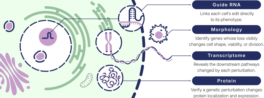
Image taken from: https://www.elementbiosciences.com/applications/crispr-screening?hsLang=en
Cytos readouts
Cell morphology (Cell Paint)
Cytos provides fluorescence-based imaging of four core cellular structures:
- Cell membrane
- Nucleus
- Actin cytoskeleton
- Mitochondria
These markers enable automated cell segmentation and extraction of morphological features, allowing researchers to investigate changes in cellular structure and phenotype following genetic perturbations or drug treatments.
Protein detection
Protein detection uses immunofluorescence-based chemistry to measure protein expression and localization at single-cell resolution.
- Detection of up to 20 protein targets (20-plex).
- Predefined protein panels or custom protein targets.
- Detection of signaling and pathway activation, including phospho-proteins.
- Intensity-based protein measurements.
Protein detection is an optional readout that can be incorporated into the different Cytos configurations.
3′ Transcriptome
Cytos uses Direct In Sample Sequencing (DISS) to capture polyadenylated RNA transcripts directly within fixed cells.
This provides an unbiased 3′ transcriptome readout, enabling researchers to investigate gene expression changes, identify affected pathways and characterize cellular responses to experimental perturbations.
Guide RNA identification
For CRISPR screening applications, Cytos enables direct sequencing of guide RNAs within individual cells.
This allows researchers to associate each genetic perturbation with its corresponding cellular phenotype, protein expression and transcriptomic response.
The platform supports screening libraries containing tens to thousands of guide RNAs simultaneously within the same sample.
Product configurations
Cytos offers three main configurations. All configurations include the core four-marker Cell Paint morphology panel, while protein detection can be added as an optional readout.
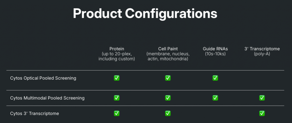
Image taken from: https://www.elementbiosciences.com/products/cytos
1. Cytos Optical Pooled Screening (OPS)
Combines guide RNA identification with cellular morphology and optional protein detection. This configuration is particularly suitable for CRISPR screens where the main objective is to link genetic perturbations to cellular phenotypes.
2. Cytos 3′ Transcriptome
Combines whole-transcriptome profiling with cellular morphology and optional protein detection. This configuration is suitable for investigating cellular responses to drug treatments or other experimental conditions without guide RNA identification.
3. Cytos Multimodal Pooled Screening (MPS)
Integrates guide RNA identification, 3′ transcriptome profiling, cellular morphology and optional protein detection. This configuration provides the most comprehensive readout, linking genetic perturbations to downstream transcriptional, protein and morphological changes within the same cell.
Applications
Cytos enables researchers to investigate cellular phenotypes and molecular responses through several complementary applications:
- CRISPR screening: Identify the effects of genetic perturbations by linking guided RNA identity to cellular morphology, protein expression and transcriptomic changes.
- Drug screening: Investigate drug responses, dose-dependent effects and potential mechanisms of action.
- Pathway analysis: Characterize changes in gene expression and protein signaling following experimental perturbations.
- Phenotypic profiling: Identify morphological changes associated with different cellular conditions or treatments.
How to start a Cytos project at NGI
- Contact us at support@ngisweden.se to discuss your experimental design and determine the most suitable Cytos configuration.
- Submit an order through the NGI order portal.
- Prepare and fix your cells on Cytos Slide Kits according to the recommended Element Biosciences sample preparation guidelines.
- Send the prepared slides to NGI for processing on the AVITI24 platform.
Sample requirements
Cytos requires cells to be prepared and fixed on dedicated Cytos Slide Kits before the screening run.
- Users are responsible for cell seeding, culturing and fixation on Cytos Slide Kits before sample submission.
- Cytos Slide Kits are available in 1-well, 12-well and 48-well configurations.
- Sample preparation and fixation must follow the Element Biosciences Cytos Sample Preparation & Optimization User Guide.
Please consult the sample submission guidelines before sending samples.
What we do with your samples
Upon arrival at NGI, the submitted slides are processed according to the selected Cytos configuration.
The slides are assembled into AVITI24 flow cells and processed using the appropriate Cytos cartridge workflow. Fluorescence-based imaging enables cell morphology characterization and, when included, protein detection. Direct In Sample Sequencing enables guide RNA and/or 3′ transcriptome detection directly within the cells.
Following the run, the instrument performs onboard analysis, including cell segmentation and guide RNA assignment when applicable.
Bioinformatics and data analysis
Further details regarding data processing, quality control reports and available downstream analysis options can be discussed when planning your project.
Further information
For detailed information about the technology, sample preparation and experimental design, please refer to the Element Biosciences Cytos documentation.
Contact: support@ngisweden.seusers get a multiQC report plus segmented data (CytoCanvas data) ready to be used for secondary analysis.
Last Updated: 18th November 2026