Visium HD 3´ – PolyA-based
NGI offers high resolution spatial transcriptomics through the 10x Genomics Visium HD 3’, which combines histology with PolyA-based transcriptomics in HD spatial context
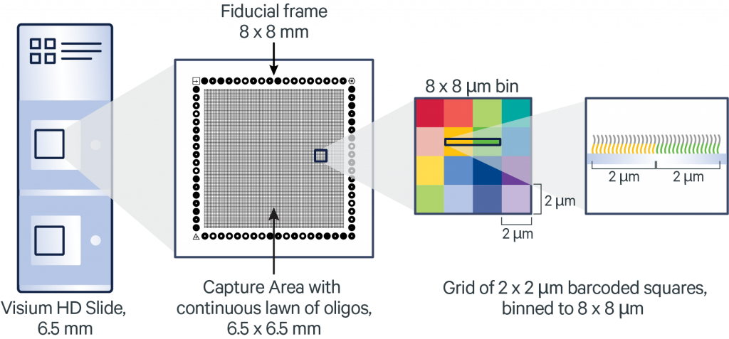
Visium HD 3’ is a spatial transcriptomics method developed by 10x Genomics that enables gene expression profiling with high spatial resolution (subcellular, up to 2 μm) from fresh frozen (FF) tissue sections placed on glass slides. Unlike the standard Visium platform, Visium HD incorporates a novel slide design and spot layout to drastically improve resolution and capture efficiency. The method captures polyadenylated mRNA transcripts, therefore being species-agnostic, enabling the detection of gene expression patterns at near single-cell level, while preserving tissue architecture.
The protocol utilises the CytAssist instrument, on four 6.5 x 6.5 mm capture area sizes. You can section the FF tissue samples in your own lab, place them on superfrost slides, and send those to us for processing. For more information about this species-agnostic spatial transcriptomics methodology, please refer to the company webpage the Visium HD 3’ Spatial Gene Expression User Guide.
Please contact us before planning your experiments for a discussion
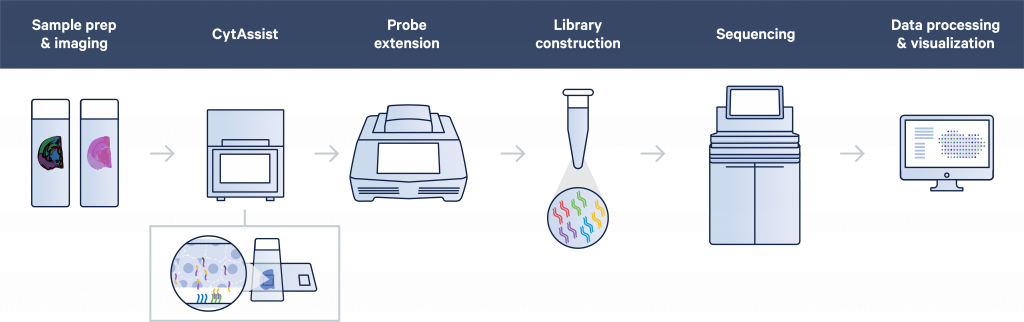
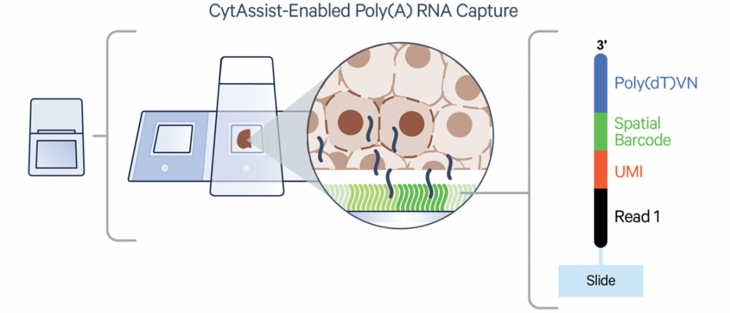
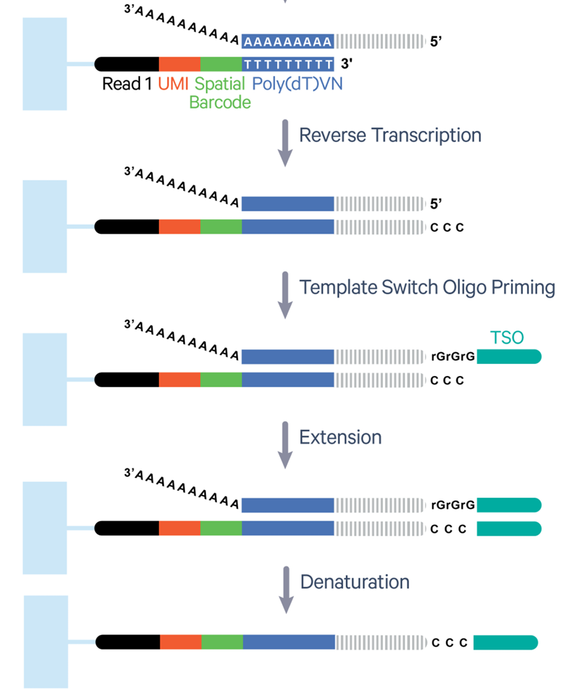
Image: The Visium HD 3’ workflow (https://www.10xgenomics.com/)
IMPORTANT! The time in between the sectioning by the user and the processing at NGI cannot exceed 4 weeks
Sample requirements
- This method relies on capturing the PolyA tail of the mRNA, therefore is a species-agnostic method and can potentially be used for any eukaryotic species.
- Currently, Visium HD 3’ is only available for 6.5 mm x 6.5 mm capture areas.
- Each kit contains a total of 4 capture areas, and this is the minimum we offer at NGI.
- Please, extract RNA from the same tissue block from which sections for Visium HD 3’ will be taken, when possible. We require you to estimate the RIN value of the extracted RNA (on a BioAnalyzer or similar), which should be greater than 7 to maximise chances of success at library preparation.
- We require you to check that the morphology of the sections is good enough for cell type annotations. This can be assessed by H&E staining of sections consecutive to the ones you submit to NGI. Please, send us the images that you obtained highlighting your region of interest, prior to sample delivery. We will use this as a reference when selecting the areas from where the RNA should be transferred and will not be able to evaluate the quality of the staining with low resolution images.
- Any staining optimisations need to be done by the user otherwise we will follow 10X Genomics’ standard protocol.
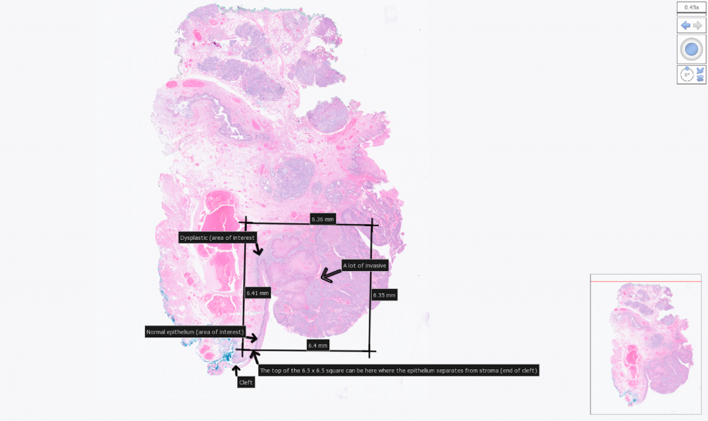
Example tissue section placement for a 6.5 mm CytAssist. Areas of interest within the section were indicated by the user with arrows.
- We require you to section and place the FF sections on coated or charged glass slides e.g VWR SuperFrost Plus Slides.
- Only one tissue section should be placed per slide!
- Recommended section thickness for most tissue types is 10 µm, but tissues from 10−20 µm are compatible with the assay.
- Sections must be placed within the specific margins described in the image below. Sections outside the margins cannot be successfully transferred. Optimise section quality and practice section placement within the allowable area before working with experimental block.
- Store the slides at -80ºC until delivery.
- Library preparation must be started within 4 weeks after sectioning so this needs to be coordinated with NGI.
- We recommend that you provide us with a back-up sample for each sample that is submitted to us. These can be returned to you upon request.
- Check the Visium HD 3’ Spatial Gene Expression User Guide for further information
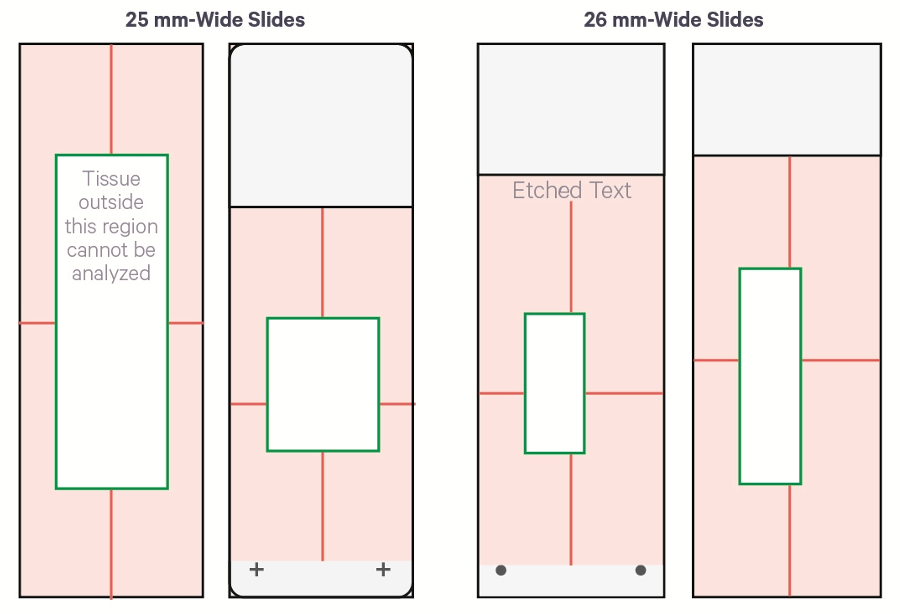

Examples of placement of sections. On the top row there examples of good placement of sections within the margins of the slides. The bottom row shows the cassette used to transfer the probes to the Visium slide.
Information we need prior to sample delivery
- Once your order has been accepted, you will receive a project Agreement. Please, read it to make sure it corresponds to your specifications and if everything looks ok, please sign it and return to us electronically.
- If the samples are from humans of other primates, a safety declaration will be sent to you along with the user agreement, which also requires to be signed.
- You will also receive a Sample Information Sheet to fill in the sample names and the RIN values derived from sections you analysed. Please send it back to us when you have the information.
- Together with these documents, we will send you slide mailers labelled with the barcode of the project. You can use these to send the slides back when ready. We will send one slide mailer per sample, which can contain also its backup sample is the case.
- You need to send us high resolution images of H&E stained sections adjacent to the samples as previously described, clearly highlighting the area of interest (see example above). We will not be able to evaluate the quality of the staining with low resolution images.
- Users are strongly encouraged to submit one backup for every sample that is submitted.
Instructions for sample labelling and delivery
- Use the pre-labelled slide mailers to send us the superfrost slides with your sections. Each slide mailer should contain a slide with your section of interest and a backup slide (if possible).
- The project-specific barcode label is a combination of your project ID (Pxxxxx) – see user agreement – and a serial number: e.g. `PxxxxxP1` for the first container. Each slide needs to be labelled with the NGI sample IDs from the Visium sample information sheet using pencil, xylene resistant stickers or markers, e.g.:
- Container `PxxxxxP1` will contain slides labelled `Pxxxxx_101`-`Pxxxxx_101-backup`
- Container `PxxxxxP2` will contain slides labelled `Pxxxxx_201`-`Pxxxxx_201-backup`
- The slide mailers must be shipped properly sealed and on dry ice. Alternatively, the user can deliver the mailers in person, always in a box with dry ice.

Example of slide container. The image shows the slide container that we will be send to the users. It has a label with the barcode corresponding to the project. Inside, they can fit a maximum of 4 slides and which should be individually labelled with the name of the sample.
What we do with your samples
Tissue sectioning is not included in our services using the CytAssist protocol for Visium HD. The FF samples will be first fixed, H&E stained, and imaged. This will be followed by the permeabilisation and transferring of the mRNAs to the Visium slide using the CytAssist instrument. RT reaction, extension will be performed on the Visium slide and the subsequent steps to add the individual indexes are carried out in reaction tubes. Last would be the sequencing and the QC of the results and the run of SpaceRanger.
Library QC and sequencing
All individual libraries will be assessed for their quality, this includes concentration measurements and estimation of average fragment lengths. The libraries will be pooled based on these results, and later sequenced on Illumina NovaSeq XPlus, depending on the sequencing depth required.
The minimum sequencing depth for Visium HD 3′ Spatial Gene Expression is 550 million read pairs per fully-covered Capture Area. We may need to adjust the sequencing setup after we know the actual tissue coverage and result of library QC. Sequencing is carried out using a 43-10-10-75 read setup: 43 cycles in read 1, two index reads with 10 cycles in each and then 75 cycles in read 2.
Bioinformatics
Sequencing data QC is handled by the NGI and runs through the 10x Genomics Space Ranger analysis pipeline. Raw data and Loupe visualisation files are delivered. You can read more about Spatial Transcriptomics analysis here.
Policy for damaged and/or misplaced sections
NGI will proceed with library preparation unless we deem user-supplied sections to be damaged, sub-optimally placed on slides, or not at a suitable thickness for the method. Therefore, it is in the best interest of users to ensure that our stated sample requirements are strictly adhered to. We strongly encouraged to submit one backup for every sample.
Signing an NGI-issued agreement for your project signifies that you accept that our staff can decide how to proceed with samples that are submitted to us. Decisions may include but are not limited to:
- Proceeding with samples that do not strictly fulfil our sample requirements but have a fair chance of yielding libraries (albeit at a lower quality than would otherwise be expected), etc.
- Charging a flat fee of SEK 5.000 as a processing fee for every back-up sample that is processed at NGI if the user has not adhered to our sample requirements for section placement on slides.
- Aborting projects comprising samples that are suboptimal for Visium.
Note that we can never guarantee any outcomes of library preparation or the preceding steps for the Visium protocol, especially if samples do not meet the criteria defined by 10x Genomics and that we outline.
Last Updated: 9th June 2026