10X Chromium Universal 3’ /5’ Gene Expression
Profiling of gene expression levels at single-cell resolution.
10X Chromium offers whole transcriptome profiling at the single cell level. In the partitions generated in the emulsion, transcripts are captured by oligos on 10X gel beads and receive both a cell-specific barcode and a UMI during reverse transcription.
Transcriptome analysis of single cells using 10X Chromium can be performed using either the 3’ or the 5’ kits. the main difference is which end of the transcript is captured onto the gel beads. For 3´ gene expression (GE), the gel beads contain poly(dT) oligos to capture the 3’ end of the transcripts whereas for the 5’ GE beads the 5’ end of the transcripts are captured by a TSO.
If you are interested in running your 3’ or 5’ GE experiment at NGI, please contact us at support@ngisweden.se.
Compatible sample types
The Chromium 3’ and 5’ GE methods are compatible with cell or nuclei suspensions from diverse species.
Sample requirements
In order to obtain high quality data from the experiment the sample should have:
- No or minimal amounts of cell debris.
- No or low levels of cell/nuclei aggregates.
- High cell viability of >80%.
- For nuclei: Intact nuclei with high-quality nuclear membranes and <5% of intact cells.
- A concentration of 700-1200 cells/nuclei per µl for a targeted recovery of 500-10 000 cells/nuclei.
- A concentration of 1300-1600 cells/nuclei per µl for a targeted recovery of 10 000-20 000 cells/nuclei.
- A maximum cell size of 30 µm. Larger cell sizes might lead to clogging of the microfluidic channels. If you have cells larger than 30 µm, it is recommended to isolate nuclei.
We always recommend performing a trial prep for new sample types before handing over any samples to NGI. The sample quality must be assessed before sample delivery. For nuclei, we recommend to assess nuclei quality at 60x in a microscope.
Extra options
Chromium 3’ and 5’ gene expression is compatible with Feature Barcoding/Cell surface protein detection.
5’ Gene expression also offers the option of Immune profiling (V(D)J analysis) for human or mouse samples, BEAM antigen repertoire mapping, as well as CRISPR analysis.
V(D)J
The Single Cell 5′ protocol offers solutions for measuring immune repertoire information and gene expression from the same cell. Profile full length (5′ UTR to constant region), paired T-cell receptor (TCR), or B-cell receptor (BCR) transcripts from the same cell.
How it works
After generating the full-length cDNA using the 10X Chromium 5’ GE kit, the 10X barcoded, full-length V(D)J segments are enriched using primers specific to the TCR or BCR constant regions. The resulting, enriched sample is used to prepare dual-indexed sequencing libraries, in which enzymatic fragmentation creates variable size fragments that collectively cover the V(D)J region.
Sample and method compatibility
Immune profiling is compatible with both cell and nuclei samples from mouse and human. If both T-cells and B-cells are expected to be present in the sample, it is possible to create libraries for both, resulting in one GE library, one BCR library and one TCR library from the same sample.
In addition, Immune profiling can be combined with feature barcode technology for cell surface protein and immune receptor mapping as well as CRISPR screening.
CRISPR
Single cell CRISPR screens can be used to study genetic regulatory networks. Using 10X Genomics’ technology CRISPR-mediated perturbations can be read out alongside a cell’s gene expression at single cell resolution.
We offer both Single Cell 5′ CRISPR Screening (GEM-X and Next-GEM) and Single Cell 3′ CRISPR Screening (Next-GEM). The choice of method will primarily depend on whether you would like to use GEM-X (recommended) or Next-GEM, and whether the assay should be combined with VDJ sequencing and/or cell surface protein detection.
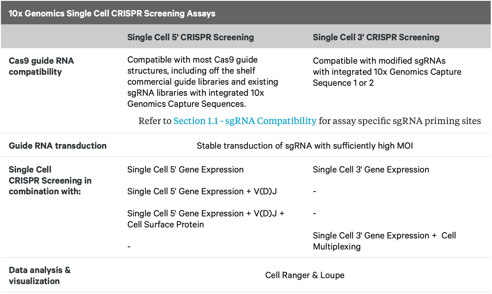
From: CG000398_ChromiumSingleCellCRISPR_Screening_Expt.PlanningGuide_Rev_C.pdf
IMPORTANT! It is crucial to test the guide efficiency and to determine the guide frequency of each sgRNA in the pool before starting any single cell experiment.
Sample requirements
Same as for 3’ and 5’ GE.
10X Genomics recommends screening 100-200 cells per targeting guide RNA and 500-1000 cells for non-targeting guide RNAs.
Guide compatibility with single cell 5′ CRISPR assay
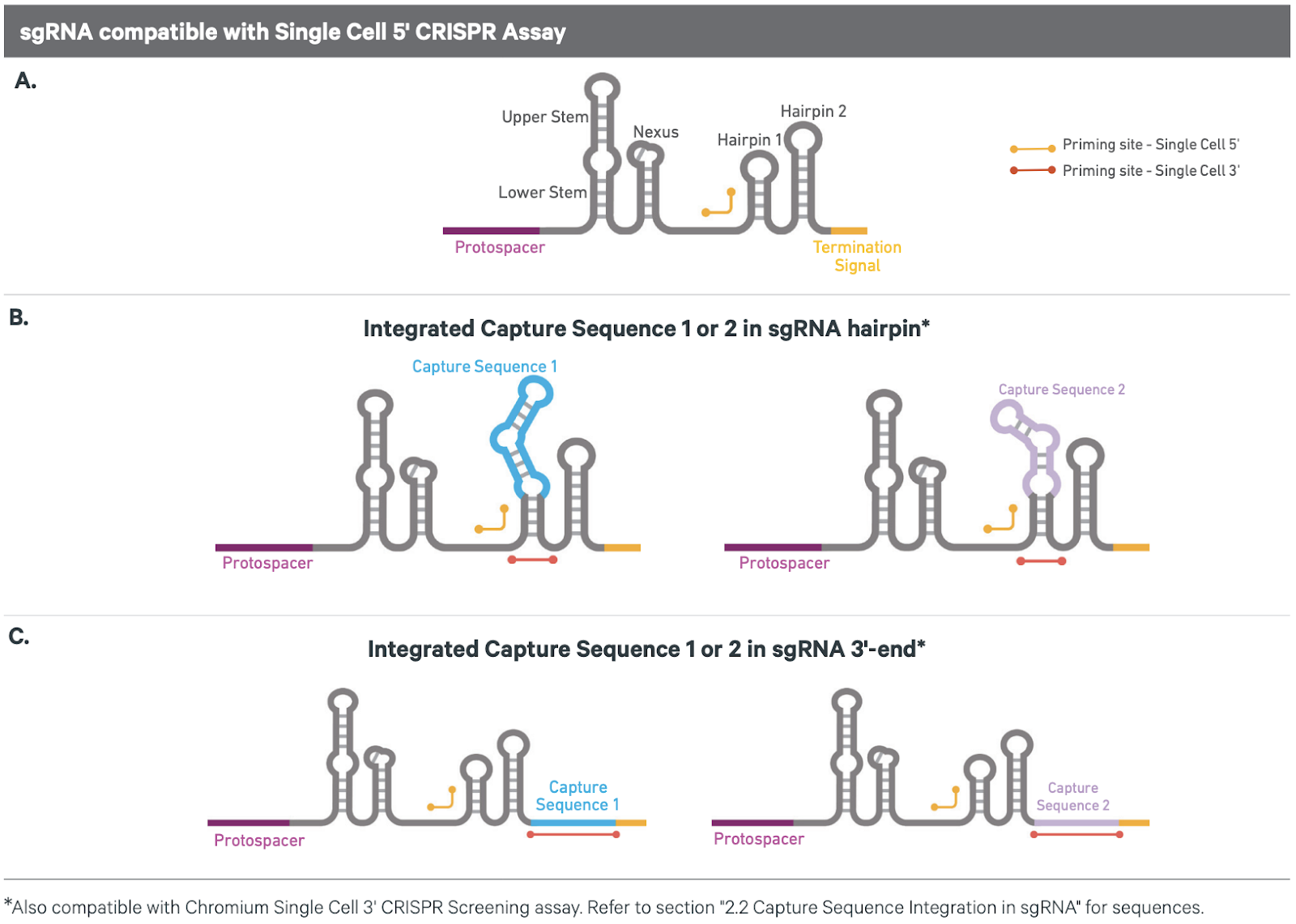
From: CG000398_ChromiumSingleCellCRISPR_Screening_Expt.PlanningGuide_Rev_C.pdf
Recommended reading: 10X Genomics CRISPR Experiment Planning Guide
Feature barcode detection
Both the 3’ GE and 5’ GE kits offer the possibility of capturing feature DNA barcodes introduced before the GEM generation. Feature barcoding can be used for multiple purposes (see table).
Feature barcoding techniques and compatibility
| Feature barcode usage | kit compatibility (GEM-X) |
| Antibody capture for cell surface protein | 3’, 5’, FLEX* |
| Sample multiplexing (“hashing”) | 3’ and 5’ |
| CRISPR guide capture | 5’ |
| Antigen Capture (BEAM) | 5’ |
How it works
After GEM generation the feature barcode is captured on the 10X gel beads and receives the same 10x barcode as the cDNA from the cell.
After pre-amplification, the sample is used both for creating the GEX library and an enriched feature barcode library. Both libraries are sequenced and the information reconciled by the common cell specific barcode, allowing for tracking presence of the feature oligo back to the specific cell and its expression pattern.
The manner of capturing the feature barcode oligo on the beads differs between the 3’ kit and the 5’ kit.
The 3’ gel beads contain, in addition to the poly(d)T for capturing RNA, capture sequence 1 and are compatible with feature barcode oligos containing the complementary sequence.
For the 5’ kit, the feature barcode oligo must contain a capture sequence complementary to the TSO on the gel bead.
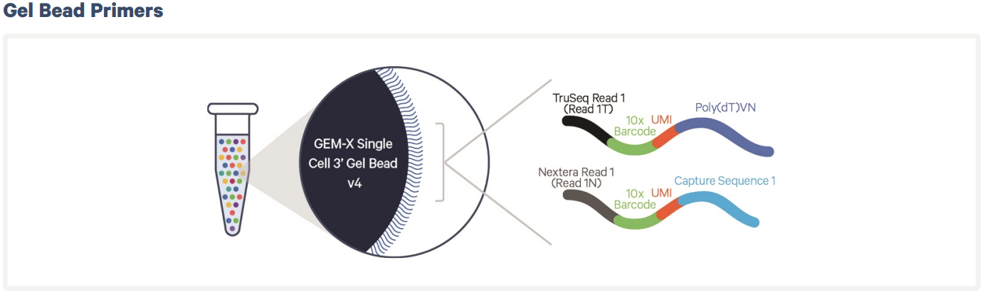
Example of feature barcode in 3’ GE. The feature barcode oligo is attached to an antibody specific to a cell surface protein. Once in the single cell emulsion the feature barcode gets captured on the bead by capture sequence 1. Transcript extension during the same step as cDNA synthesis leads to incorporation of the cell specific 10X barcode. In the 5’ kit the feature oligo gets captured by the TSO. Adapted from: https://cdn.10xgenomics.com/image/upload/v1710230668/support-documents/CG000732_ChromiumGEM-X_SingleCell3_ReagentKitsv4_CellSurfaceProtein_UserGuide_RevA.pdf
Cell surface protein
If the feature barcode oligo is coupled to an antibody specific for a cell surface protein of interest, including it in the 10x GE experiment will allow for distinguishing cells in the sample that express this particular protein and connect it with the GE profile.
Sample multiplexing using antibodies
Labeling cells with antibody-oligonucleotide conjugates can also be used as a means for sample multiplexing (also called “hashing”). For such experiments, the different cell samples are labeled with antibodies specific for a common cell surface antigen conjugated to oligos with different barcodes before the 10x experiment. The sequences from the feature barcode library (or HTO library) are used for demultiplexing the samples after sequencing.
In addition to allowing for combining multiple samples in the same 10x reaction, the same technique can be used to increase the number of cells in a single reaction by aiding in identification of multiplets. If a single cell suspension is split into 2 or more tubes and labeled with different antibody-oligonucleotide conjugates, droplets that receive more than 1 cell can be distinguished by the presence of more than 1 feature barcode. Please note that this only allows the identification and filtering of the multiplets, it cannot be used to resolve the data from the individual cells in the multiple droplet. Please note that using feature barcoding for hashing purposes is not officially supported by 10X Genomics.
Recommended antibodies
10x fully supports cell surface protein detection using the Totalseq B or Totalseq-C antibody oligonucleotide conjugates from Biolegend. Use of Totalseq-A is no longer officially supported by 10x and therefore no longer offered by NGI.
Totalseq-B is compatible with the 3’ GE kit and Totalseq-C is compatible with the 5’ GE kit.
NGI normally has a number of Totalseq-B and -C conjugates for sample multiplexing of human and mouse cells, and offers users to buy aliquots. For ordering other compatible conjugates or designing custome conjugates, we recommend contacting Biolegend.
It is also possible to conjugate oligos to other antibodies of interest. For more information please have a look at the this 10x support page.
Please note that users are responsible for optimising the conditions for antibody labeling that is performed before delivering the sample to NGI. Please refer to the 10x protocol Cell Surface Protein Labeling for Single Cell RNA Sequencing Protocols with Feature Barcode technology.
We recommend testing the protocol to generate your antibody-labelled single cell suspension before scheduling your run.
Antigen Capture – BEAM
The Chromium Single Cell 5′ Barcode Enabled Antigen Mapping (BEAM) protocol makes it possible to map antigen-specificity of B or T cells while simultaneously obtaining cell surface gene expression profiles and immune receptor sequences for the same cell.
In BEAM the antigens of interest are labeled and conjugated to feature barcode oligos (the “BEAM conjugate”) and introduced to the cell suspension. The population of immune receptors specific to the antigen bind the conjugate. The cells are also stained with Immunecell panels of labeled antibodies, allowing for FACS sorting of antigen specific B or T-cells.
The cells are then processed using the 10X Chromium 5’ kit, generating a GE library, an antigen capture library and a V(D)J library for sequencing. The method is also compatible with cell surface protein detection.
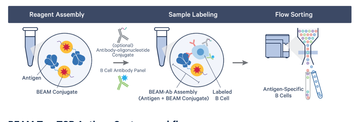
Figure outlining the principle of BEAM B-cell isolation. For T-cells the BEAM conjugate includes MHC monomers and the antigenic peptide must first be mixed with the BEAM-conjugates since the TCR only recognizes peptides in the context of the MHC. Image from https://www.10xgenomics.com/support/software/cell-ranger/latest/getting-started/cr-5p-what-is-antigen-capture
On-Chip multiplexing (OCM)
The On-Chip-Multiplexing (OCM) assay offers the possibility to multiplex your single cell experiments on the GEM-X chip, without requiring any extra cell-tagging steps. Samples from 4 different wells, carrying barcoded gel beads, are co-partitioned and collected in the same recovery well. Co-partitioned samples are demultiplexed computationally after sequencing.
How it works
Samples from 4 individual wells, containing barcoded gel beads, are co-partitioned in one collecting well. In total, 8 samples can be multiplexed and generate 2 pooled samples (each pooled sample contains 4 samples). Each individual sample out of the four in the pool, will contribute to the pooled sample with 5000 cells, resulting in a co-partitioned sample in the collecting well that will contain 20000 cells.
Advantages of the OCM assay
- Allows multiplexing of up to 4 samples per reaction, each up to 5000 cells without any additional steps. For example:
- Sample types with limitied cell numbers.
- pooling of technical replicates
- More cost efficient when analysing samples with target of <5000 cells.
- The OCM assay is compatible with the same multiomic capabilities offered for 3’ or 5’ workflows, without requiring additional steps.
Disadvantages of the OCM assay
- Higher multiplet rate than 10X genomics standard 3′ or 5′ assays. For example 7.6% multiplets if analysing 5000 cells per sample

Figure from 10X website.
Starting a single cell Universal 3’ or 5’ GE project with NGI
- Contact us at support@ngisweden.se to discuss your project.
- Read our current user guidelines here (v14).
- If possible – run a pilot for the sample preparation to ensure that it yields high quality cell suspensions/nuclei.
- Submit an order via the NGI order portal, using the “Single-cell library preparation and sequencing” order form (you’ll need to make an account first, if you do not have one already).
- Book a day/days to bring your samples to us.
Information we need prior to sample delivery
- A signed agreement and filled-in sample information sheet (both provided to you when we have processed your submitted order).
- A filled-in safety declaration that is sent to you along with the user agreement (if applicable).
What we do with your samples
Upon delivery of the samples to NGI, the responsible lab staff will count the cells and check the quality of the cell/nuclei suspension before you leave.
Should the sample be sub-optimal and you still wish to continue with the experiment, we will ask you to confirm this in writing. In such a situation you will be charged the cost of the 10x reactions regardless of the outcome. If you decide not to continue, there will be no charge unless a kit was purchased specifically for you (communicated to you before setting up your project).
All custom sample manipulation such as labeling, multiplexing etc must be performed by the researcher prior to delivery of the sample. The delivered sample should be ready to load on the 10X Chromium instrument.
Library preparation
1. GEM generation, barcoding and RT
The samples are loaded on the Chromium X controller for GEM generation and cDNA synthesis inside the GEMs. During cDNA synthesis, a cell-specific barcode is incorporated into the full-length cDNA inside the GEMs.
2. Post GEM-RT cleanup, cDNA amplification and cDNA QC
The emulsion is broken by the addition of a recovery agent and the first-strand cDNA is purified before cDNA pre-amplification. The amplified cDNA is purified and the quality is checked by BioAnalyzer.
3. cDNA purification and library preparation
The cDNA is enzymatically fragmented and indexed to produce the final sequencing library.
4. Library QC and Sequencing
The library is QCed by Qubit, BioAnalyzer and qPCR before pooling for sequencing.
Bioinformatics
Basic bioinformatic analysis using the CellRanger software from 10X Genomics. Your data will be delivered to you through the DDS online delivery system.
Last Updated: 1st June 2026